Nanoscale imaging of clinical specimens using pathology-optimized expansion microscopy
[Publisher Link] [Local Copy]
Zhao Y*, Bucur O*, Irshad H, Chen F, Weins A, Stancu AL, Oh EY, DiStasio M, Torous V, Glass B, Stillman IE, Schnitt SJ, Beck AH**, Boyden ES** (2017) Nanoscale imaging of clinical specimens using pathology-optimized expansion microscopy, Nature Biotechnology 35(8):757-764. (*, co-first authors; **, co-corresponding authors)
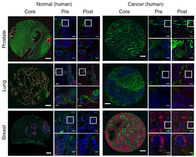
Expansion microscopy (ExM), a method for improving the resolution of light microscopy by physically expanding a specimen, has not been applied to clinical tissue samples. Here we report a clinically optimized form of ExM that supports nanoscale imaging of human tissue specimens that have been fixed with formalin, embedded in paraffin, stained with hematoxylin and eosin, and/or fresh frozen. The method, which we call expansion pathology (ExPath), converts clinical samples into an ExM-compatible state, then applies an ExM protocol with protein anchoring and mechanical homogenization steps optimized for clinical samples. ExPath enables ~70-nm-resolution imaging of diverse biomolecules in intact tissues using conventional diffraction-limited microscopes and standard antibody and fluorescent DNA in situ hybridization reagents. We use ExPath for optical diagnosis of kidney minimal-change disease, a process that previously required electron microscopy, and we demonstrate high-fidelity computational discrimination between early breast neoplastic lesions for which pathologists often disagree in classification. ExPath may enable the routine use of nanoscale imaging in pathology and clinical research.
Resources associated with this Publication:
[Expansion microscopy: physical magnification with nanoscale precision]