Expansion Microscopy of Zebrafish for Neuroscience and Developmental Biology Studies
[Publisher Link] [Local Copy]
Freifeld L, Odstrcil I, Förster D, Ramirez A, Gagnon JA, Randlett O, Costa EK, Asano S, Celiker OT, Gao R, Martin-Alarcon DA, Reginato P, Dick C, Chen L, Schoppik D, Engert F, Baier H, Boyden ES (2017) Expansion microscopy of zebrafish for neuroscience and developmental biology studies, Proceedings of the National Academy of Sciences 114(50):E10799-E10808.
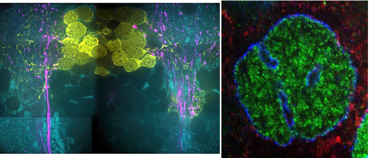
Expansion microscopy (ExM) allows scalable imaging of preserved 3D biological specimens with nanoscale resolution on fast diffraction-limited microscopes. Here, we explore the utility of ExM in the larval and embryonic zebrafish, an important model organism for the study of neuroscience and development. Regarding neuroscience, we found that ExM enabled the tracing of fine processes of radial glia, which are not resolvable with diffraction-limited microscopy. ExM further resolved putative synaptic connections, as well as molecular differences between densely packed synapses. Finally, ExM could resolve subsynaptic protein organization, such as ring-like structures composed of glycine receptors. Regarding development, we used ExM to characterize the shapes of nuclear invaginations and channels, and to visualize cytoskeletal proteins nearby. We detected nuclear invagination channels at late prophase and telophase, potentially suggesting roles for such channels in cell division. Thus, ExM of the larval and embryonic zebrafish may enable systematic studies of how molecular components are configured in multiple contexts of interest to neuroscience and developmental biology.