Expansion microscopy: development and neuroscience applications
[Publisher Link] [Local Copy]
Karagiannis ED, Boyden ES (2018) Expansion microscopy: development and neuroscience applications, Current Opinion in Neurobiology 50:56-63.
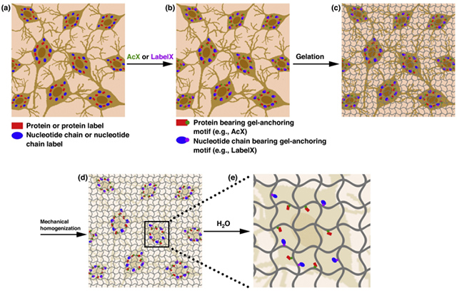
Many neuroscience questions center around understanding how the molecules and wiring in neural circuits mechanistically yield behavioral functions, or go awry in disease states. However, mapping the molecules and wiring of neurons across the large scales of neural circuits has posed a great challenge. We recently developed expansion microscopy (ExM), a process in which we physically magnify biological specimens such as brain circuits. We synthesize throughout preserved brain specimens a dense, even mesh of a swellable polymer such as sodium polyacrylate, anchoring key biomolecules such as proteins and nucleic acids to the polymer. After mechanical homogenization of the specimen-polymer composite, we add water, and the polymer swells, pulling biomolecules apart. Due to the larger separation between molecules, ordinary microscopes can then perform nanoscale resolution imaging. We here review the ExM technology as well as applications to the mapping of synapses, cells, and circuits, including deployment in species such as Drosophila, mouse, non-human primate, and human.
Resources associated with this Publication:
[Expansion microscopy: physical magnification with nanoscale precision]