Expansion Microscopy for Beginners: Visualizing Microtubules in Expanded Cultured HeLa Cells
[Publisher Link] [Local Copy]
Zhang C*, Kang JS*, Asano SM, Gao R, Boyden ES (2020) Expansion Microscopy for Beginners: Visualizing Microtubules in Expanded Cultured HeLa Cells, Current Protocols in Neuroscience 92(1):e96. (*, contributed equally)
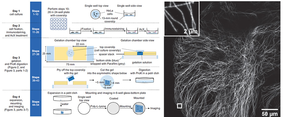
Expansion microscopy (ExM) is a technique that physically expands preserved cells and tissues before microscope imaging, so that conventional diffraction-limited microscopes can perform nanoscale-resolution imaging. In ExM, biomolecules or their markers are linked to a dense, swellable gel network synthesized throughout a specimen. Mechanical homogenization of the sample (e.g., by protease digestion) and the addition of water enable isotropic swelling of the gel, so that the relative positions of biomolecules are preserved. We previously presented ExM protocols for analyzing proteins and RNAs in cells and tissues. Here we describe a cookbook-style ExM protocol for expanding cultured HeLa cells with immunostained microtubules, aimed to help newcomers familiarize themselves with the experimental setups and skills required to successfully perform ExM. Our aim is to help beginners, or students in a wet-lab classroom setting, learn all the key steps of ExM.
Resources associated with this Publication:
[Expansion microscopy: physical magnification with nanoscale precision]