Expansion microscopy of C. elegans
[Publisher Link] [Local Copy]
Yu CJ, Barry NC, Wassie AT, Sinha A, Bhattacharya A, Asano S, Zhang C, Chen F, Hobert O, Goodman MB, Haspel G, Boyden ES (2020) Expansion microscopy of C. elegans, eLife 9:e46249.
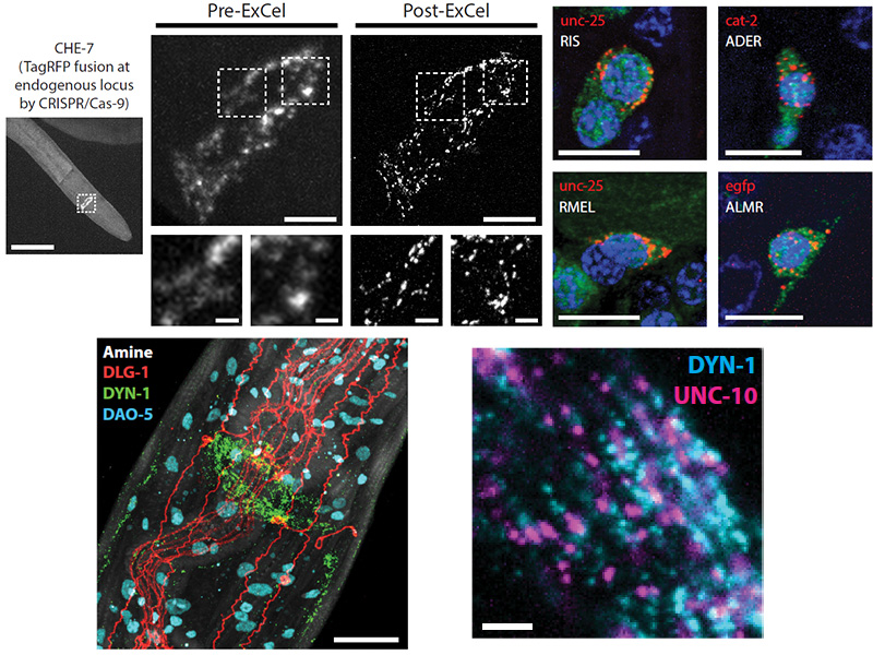
We recently developed expansion microscopy (ExM), which achieves nanoscale-precise imaging of specimens at ~70 nm resolution (with ~4.5x linear expansion) by isotropic swelling of chemically processed, hydrogel-embedded tissue. ExM of C. elegans is challenged by its cuticle, which is stiff and impermeable to antibodies. Here we present a strategy, expansion of C. elegans (ExCel), to expand fixed, intact C. elegans. ExCel enables simultaneous readout of fluorescent proteins, RNA, DNA location, and anatomical structures at resolutions of ~65–75 nm (3.3–3.8x linear expansion). We also developed epitope-preserving ExCel, which enables imaging of endogenous proteins stained by antibodies, and iterative ExCel, which enables imaging of fluorescent proteins after 20x linear expansion. We demonstrate the utility of the ExCel toolbox for mapping synaptic proteins, for identifying previously unreported proteins at cell junctions, and for gene expression analysis in multiple individual neurons of the same animal.
Resources associated with this Publication:
[Expansion microscopy: physical magnification with nanoscale precision]